Month: July 2025

  • Catching ghosts: what environmental DNA can (and can’t yet) do for biosecurity surveillance of biting midges

    Catching ghosts: what environmental DNA can (and can’t yet) do for biosecurity surveillance of biting midges

    Biting midges of the genus Culicoides are tiny, often just 1–3 mm long, but their impact on animal health and agricultural economies can be vast. They vector more than 50 viruses of veterinary concern, including bluetongue and African horse sickness. New Zealand remains free of Culicoides and the diseases they transmit, and it runs a national surveillance programme to keep it that way. This article shares a study that shows advancements in the utilisation of DNA-based tools, specifically DNA metabarcoding of bulk insect samples and environmental DNA (eDNA) recovered from trap fluids to make surveillance faster, more scalable and no less reliable.

    Early detection is everything in biosecurity. If Culicoides arrived and established silently, the first sign might be an animal disease outbreak that is both costly and difficult to contain. Yet scanning 15,000–20,000 insects a year by eye is neither efficient nor easily expandable. Molecular approaches promise to read the genetic “fingerprints” of all organisms in a mixed sample in one go, potentially flagging rare or unexpected species that taxonomists might miss.

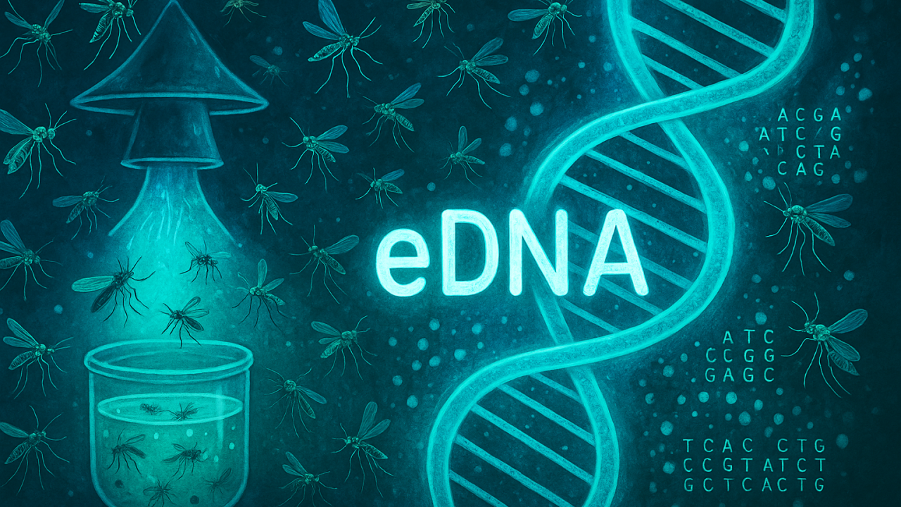
    A brief primer on metabarcoding and eDNA

    Metabarcoding is a high-throughput method that uses universal or semi‑universal genetic primers to amplify barcode regions (here, COI) from all organisms present in a mixed sample, which are then sequenced en masse. Bioinformatic clustering of the reads into operational taxonomic units (OTUs) allows rapid estimation of which taxa are present. Environmental DNA (eDNA) refers to the genetic material shed by organisms into their surroundings—water, soil, air, or, in the case of this study, the ethanol inside a light trap—so that it can be captured without handling the organism itself. Both approaches can dramatically reduce the time to screen complex samples, but both come with biases linked to primer choice, DNA degradation, biomass differences and amplification stochasticity.

    What the team actually did

    The researchers analysed 38 trap samples collected weekly over nine to ten weeks in 2020 from four cattle farms in different North Island districts: Morrinsville, Okaihau, Warkworth and Whakatāne. Each site deployed green LED light traps baited with carbon dioxide and octenol to attract midges and other flying insects. The traps were filled with ethanol to preserve the catch. Every sample was first processed morphologically: insects were counted, and any members of the midge family Ceratopogonidae were recorded. As Culicoides does not occur in New Zealand, native Ceratopogonidae served as a proxy “target group” to test how well the DNA methods could recover what morphology saw and spot what morphology might have missed.

    Two parallel molecular routes were then compared. In one, the entire insect catch was homogenised (destroying the specimens) and DNA extracted from the tissue mixture. In the other, DNA was filtered from the ethanol preservative—the eDNA that insects shed into the fluid—leaving the intact specimens available for later taxonomic work if needed. Both DNA types were amplified using two commonly used mitochondrial cytochrome oxidase I (COI) primer pairs (LCO1490/HCO2198 and mlCOIintF/jgHCO2198) and sequenced on an Illumina MiSeq platform. An in‑house Ceratopogonidae COI reference library, supplemented by public databases, supported species assignment.

    What the microscope found

    Across the 38 trap samples, a total of 45,745 insects were counted. No exotic Culicoides were detected, but native Ceratopogonidae were present in 22 traps (58%), albeit at low abundance—just 114 individuals overall, representing a mere 0.25% of total catch. Most of those were from Whakatāne, where Ceratopogonidae appeared in every sample. These figures served as the benchmark for evaluating molecular performance.

    Bulk metabarcoding outperformed eDNA for target detection

    When it came to reproducing the morphological verdict on whether Ceratopogonidae were present or absent, metabarcoding of homogenised bulk samples was consistently more accurate than metabarcoding of eDNA from the trap fluid. Using the classic LCO1490/HCO2198 primers, bulk metabarcoding achieved an overall detection accuracy of 81.94%, compared with 68.42% for the ethanol-derived eDNA. With the mlCOIintF/jgHCO2198 primers, bulk metabarcoding again reached roughly the same accuracy (81.58%), while eDNA accuracy dropped to 55.26%. In short, across both primer sets, the bulk approach surpassed the 80% mark, whereas eDNA lagged behind.

    False negatives—cases where Ceratopogonidae were seen under the microscope but not detected by sequencing—were the main reason eDNA underperformed. The team also observed a few ‘false positives’, where metabarcoding detected Ceratopogonidae that morphology did not; these could reflect genuinely missed specimens, tiny fragments invisible to the taxonomist, or technical artefacts such as low‑level index “cross‑talk” among samples during sequencing. Importantly, metabarcoding (both eDNA and bulk) did sometimes rescue detections that morphology missed, reminding us that the microscope is not infallible either.

    Why did eDNA struggle?

    The biology and physics are not in eDNA’s favour here. Ceratopogonidae are extremely small. Small-bodied insects contribute little biomass and therefore little DNA to the preservative ethanol, especially compared with the large moths and flies that dominate trap catches. eDNA is also not evenly distributed through the fluid and can degrade quickly. Even when present, amplification biases during PCR can down‑weight scarce templates further, particularly with broad “universal” primers. Together, these factors make it hard for eDNA metabarcoding to reliably pick up low‑abundance, tiny-bodied targets amid a noisy community background—at least with generalist primers and the workflows used here.

    Primer choice shaped the community picture more than sample type

    Although eDNA was weaker for yes/no detection of Ceratopogonidae, both molecular approaches painted broadly similar pictures of the overall insect communities in each trap. Non-metric multidimensional scaling showed that ethanol and bulk samples from the same trap usually clustered together, meaning they recovered comparable community structure. What did make a bigger difference was the primer pair. LCO1490/HCO2198 skewed strongly towards Lepidoptera, whereas mlCOIintF/jgHCO2198 yielded a more even spread across orders such as Lepidoptera, Diptera and Trichoptera. This reinforces a central truth of metabarcoding: your primers define your window on biodiversity.

    So, what value does eDNA bring?

    Despite its lower sensitivity for the specific, tiny-bodied target group tested here, eDNA retains several important advantages. It is non‑destructive, allowing taxonomists to re‑examine the original specimens. It is operationally simpler—filter the fluid, extract DNA, and you can process far more samples more quickly than microscopy allows. It produces community‑level data that align well with bulk metabarcoding, so it can still track spatial and temporal shifts in insect assemblages at scale. Crucially, eDNA’s performance could be markedly improved if the aim is targeted detection rather than broad community profiling—for example, by switching from metabarcoding to quantitative PCR (qPCR) or digital PCR assays that lock onto Culicoides‑specific markers with much higher sensitivity. In other words, use eDNA metabarcoding to understand the community and screen widely, then deploy species‑specific assays to decisively confirm or rule out incursions.

    Practical implications for biosecurity programmes

    For routine, rapid, early‑warning surveillance of unwanted midges, bulk-sample metabarcoding currently delivers the best balance of accuracy and throughput when using universal COI markers. Where preserving specimens is essential, an eDNA-first workflow could facilitate triage of samples: rapidly screen trap fluids to flag suspect traps, then prioritise those specimens for either bulk metabarcoding, species-specific qPCR, or morphological confirmation. Programmes should also invest in well-curated, regionally comprehensive reference libraries (the in-house Ceratopogonidae database used in this study was invaluable) and in validating primer sets against the taxa of greatest concern.

    Conclusion

    This study offers a measured, practice‑ready message. If your immediate need is to know, with high confidence, whether a delicate, low‑abundance target like Ceratopogonidae is present in a trap, homogenised bulk metabarcoding with carefully chosen primers currently does the job better than ethanol‑derived eDNA. But if your goal is to scale surveillance, keep specimens intact, and track whole insect communities efficiently, eDNA metabarcoding of trap fluids is already useful, and with species‑specific assays layered on top, it could become a powerful, non‑destructive frontline tool for early detection.

  • Detecting Parasitic Mites (Varroa destructor) in Honey Bee Hives using Environmental DNA

    Detecting Parasitic Mites (Varroa destructor) in Honey Bee Hives using Environmental DNA

    The health of honey bee populations underpins both global agriculture and ecological resilience. Among the many threats facing bees, the parasitic mite Varroa destructor has become a particular focus of international concern. This tiny yet devastating pest weakens colonies, transmits viruses, and contributes to significant losses in managed bee populations worldwide.

    The recent arrival of Varroa destructor in Australia, a country that had previously remained free of the mite, has underscored the urgent need for effective detection and biosecurity measures. Timely identification of infestations is critical for protecting both agricultural productivity and biodiversity. In this context, a promising innovation is emerging: the use of environmental DNA (eDNA) as a tool for monitoring V. destructor.

    This article shares recent research that investigates the application of eDNA methods for detecting Varroa destructor within honey bee hives, offering a complementary approach to traditional techniques. The findings suggest that eDNA-based detection could enhance surveillance, support early interventions, and contribute to safeguarding pollinator health globally.

    Why Early Detection Matters

    Honey bees play an essential role as pollinators of both crops and wild plants. Their decline poses risks not just to agricultural yields, but to the integrity of ecosystems more broadly. The spread of Varroa destructor has exacerbated this challenge. By feeding on bees and facilitating the transmission of pathogens, the mite undermines colony health, disrupts pollination services, and amplifies the vulnerability of already stressed bee populations.

    Traditional methods for detecting V. destructor, such as alcohol washes, are widely used by beekeepers but involve handling bees directly, which can disturb colonies and require considerable effort and expertise. These approaches are reliable but not ideally suited for frequent, large-scale surveillance, particularly when rapid response is needed.

    Environmental DNA: A New Approach

    Environmental DNA refers to genetic material that organisms shed into their surroundings, through skin, saliva, faeces or, in this case, hive debris and honey. By sampling and analysing this DNA, researchers can detect species without needing to observe or capture them directly.

    eDNA methods offer several distinct advantages. They are non-invasive, reducing the need to handle bees and disrupt colonies. The techniques are also highly sensitive, capable of detecting low levels of pest DNA that might indicate an incipient infestation. Moreover, eDNA sampling is scalable, as it can be employed across multiple sites relatively quickly and without the need for extensive specialist training, making it attractive to both beekeepers and biosecurity authorities.

    Developing the Molecular Assay

    At the heart of this study was the development of a species-specific quantitative PCR (qPCR) assay, designed to target the mitochondrial cytochrome oxidase (cox1) gene of Varroa destructor. This gene provides a unique signature, enabling precise identification of mite DNA even amidst the complex biological material present in a hive.

    To ensure reliability, the assay was rigorously tested against DNA from closely related mite species and honey bee hosts. This step was essential for confirming specificity and avoiding false positives. Optimisation of the assay parameters included careful calibration of primer and probe concentrations to maximise sensitivity.

    All assays were performed using a modern real-time PCR platform, ensuring robust and reproducible detection.

    Sampling Inside the Hive

    The research deployed a sampling strategy designed to capture eDNA from multiple sources within honey bee colonies. Honey, in particular, was recognised as a valuable substrate. Collected from capped combs using sterile techniques, honey provides a medium where mite DNA can accumulate over time, effectively recording the history of infestation.

    Swabs were taken from hive entrances and brood frames, targeting surfaces where mites or their traces might be deposited by foraging and nursing bees. To ensure accuracy, negative control samples were collected at each sampling site to monitor for contamination.

    Comparing eDNA with Traditional Techniques

    To evaluate the effectiveness of eDNA detection, the researchers compared their findings with those obtained through the alcohol wash method. In this conventional approach, around 300 adult bees are immersed in ethanol, dislodging mites for counting. While robust, this method is inherently invasive and labour-intensive.

    Statistical analyses, including McNemar’s Chi-square test, were used to assess the agreement between the two approaches, providing a quantitative measure of eDNA’s performance relative to established practice.

    Key Findings

    The study revealed that eDNA methods can offer a highly sensitive means of detecting Varroa destructor. Among the different sample types, honey consistently yielded the highest detection rates, with sensitivity measured at 91%. This outperformed swabs from hive entrances and brood frames, whose sensitivities were considerably lower.

    Importantly, the number of mite DNA copies detected in honey samples increased with infestation levels, suggesting that eDNA quantification could offer insights not only into presence or absence, but also into the scale of infestation. Such information is valuable for beekeepers aiming to tailor interventions appropriately.

    However, the study also noted limitations. Detection rates declined at very low infestation levels (below 1%), indicating that eDNA sensitivity is diminished when mite populations are just beginning to establish. This finding highlights the need for further optimisation, especially if eDNA is to be deployed as an early warning system.

    The results highlighted that while swabs from hive entrances and brood frames provided valid supplementary data, they lacked the consistency shown by honey samples. A combined approach, drawing on both honey sampling and surface swabbing, may prove to be the most effective.

    Implications for Biosecurity and Beekeeping

    The potential applications of eDNA techniques in biosecurity surveillance are significant. Their non-invasive nature reduces stress on bee colonies. At the same time, their scalability allows for broader monitoring coverage—qualities that are particularly valuable when surveillance needs to be expanded rapidly, such as during an incursion into previously mite-free regions.

    By complementing, rather than replacing, traditional methods, eDNA detection can enhance the overall surveillance toolkit available to beekeepers and regulators. Alcohol washes will remain important for estimating infestation intensity within colonies, but eDNA provides an additional, sensitive means for early detection.

    For Australia and other regions still striving to keep Varroa destructor at bay, eDNA offers a means of detecting incursions promptly, buying time for containment and eradication efforts before the mite becomes established.

    Looking Ahead

    This research opens the door to further exploration of how eDNA methods can contribute to supporting pollinator health. Collaboration will be key: researchers, beekeepers, biosecurity authorities and policymakers will need to work together to ensure that eDNA tools are developed, validated and deployed effectively. Future work could focus on improving sampling protocols, investigating the persistence of mite DNA in hive environments, and extending the approach to monitor other pests and pathogens relevant to honey bee health.

    Pollinator health is under increasing pressure. Innovations such as these represent not just technical progress but a vital contribution to securing the future of both food production and ecological stability.

  • Introduction to Environmental DNA for Conservation

    📍Venue:University of Nairobi, Chiromo Campus
    📅 Date:15 August 2025
    Time:9:00 AM – 1:00 PM EAT

    Description


    This foundational masterclass offers an accessible entry point into the science and application of environmental DNA (eDNA) for biodiversity conservation. Designed for researchers, students, conservation practitioners, and policymakers, the session explores how eDNA is transforming species monitoring and ecosystem assessment—particularly in contexts where traditional methods are costly, invasive, or ineffective.

    Key Highlights

    • 🎓 No prior molecular biology background required
    • 🧪 Includes a live demonstration of eDNA collection
    • 🌍 Focus on real-world conservation use cases from Kenya and East Africa
    • 🤝 Networking session with eDNA professionals and project leads
    • 📝 Certificate of participation available